Developing ultrasound systems for better imaging and cancer treatment
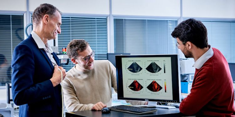
Ultrasonic embedded systems research, by Professor Steve Freear and colleagues, has been a key factor in recent microbubbles research for better cardiac imaging and chemotherapy drug release.
Embedded systems are in most of the electronics we use every day, from phones to washing machines. They are smaller versions of desktop computers but control just one function and they are power efficient and low energy. They use FPGA technology – a blank silicon block on which a code is designed and written in Verilog language for a particular function.
No other group in the UK is building their own “in-house” designed hardware – from transducers and all the electronics - and then adapting commercial FPGA tech to design the chips.
The strength of the team at Leeds is their patented hardware research platform (Ultrasonic Array Research Platform, UARP). No other group in the UK is building their own “in-house” designed hardware – from transducers and all the electronics - and then adapting commercial FPGA tech to design the chips to process multi-channel data from a wide range of applications.
Research by Professor Steve Freear and colleagues, has been a key factor in recent microbubbles research for better cardiac imaging and chemotherapy drug release
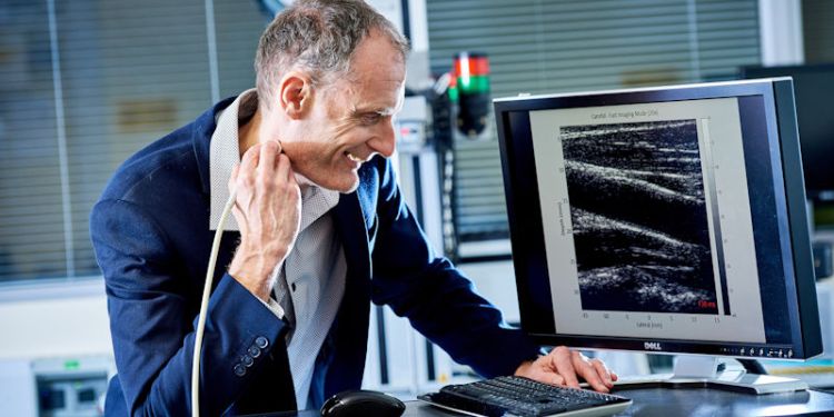
Application of UARP tech has been crucial in microbubbles research for better cardiac imaging. Microbubbles, when introduced into the heart, can be used to enhance an ultrasound image.
“The vibration of the bubbles creates a strong scatter to enhance the ultrasound image. Here the FPGA technology is crucial, as a whole DVD worth of data can be created in a few seconds by the parallel channels,” said Steve. “It gives a high-frame-rate image of the contracting heart muscle, and can slow the images down frame by frame to look at detailed flow of blood through the heart, which gives information on leaking valves or holes between chambers.”
It gives a high-frame-rate image of the contracting heart muscle, and can slow the images down frame by frame to look at detailed flow of blood through the heart, which gives information on leaking valves or holes between chambers
There are over 1 million ultrasound scans in the UK every year and the results can affect the diagnosis and medical pathway followed.
The microbubbles technology is also being adapted for use in chemotherapy for colorectal cancer with Dr Louise Coletta from the Faculty of Medicine and Health. By encapsulating the chemotherapy drugs in microbubbles and then targeting them specifically at tumour areas, it could help reduce side effects.
The pressure of the ultrasound is increased to burst the bubbles and release the drug. The system developed by Professor Freear’s team can switch easily between ultrasound imaging and the therapeutic release of drugs. This technology has received follow on funding from UK research council EPSRC and the Medicines Discovery Catapult fund. The team are now replicating studies to provide confidence in their results and the next step is licencing the process, followed by clinical trials within a couple of years.
Steve divides time between medical applications and industrial work. The two areas support each other in developing the technology.
Contact us
If you would like to discuss this area of research in more detail, please contact Professor Steve Freear.