Research project
BonE-GraphT
- Start date: 1 July 2017
- End date: 30 June 2019
- Funder: EU Horizon 2020
- Value: Overall €195 454,80 and EU contribution €195 454,80
- Primary investigator: Professor Animesh Jha
- Co-investigators: Marie-Curie Fellow Dr Chiranjeevi Maddi
Grant Agreement ID
752297

The BonE-GraphT aimed to develop a new approach, based on bone template engineering using which the entire load-bearing structure might be possible to regenerate for replacing the damaged bone. It aimed to align with the tissue-engineering for in-theatre use in order to meet the demand in treating damaged load-bearing long bones (e.g. tibia femur) in trauma, osteoporotic bones and other bone defects.
Bone is a regenerative tissue, which has the potential for healing provided the following physiological conditions are met, and these are:
i) supporting angiogenesis for carrying nutrients for tissue restoration;
ii) provide environment osteogenesis leading formation of hard minerals by using the growth factors and bone morphogenic proteins, available in situ;
iii) support mechanical function by load transmission defects are quite prevalent globally and can be the result of genetic, metabolic and pathological causes or trauma in human beings. The absence of one or more factors become apparent when a damaged bone stops healing after surgery. During healing process, the overall osteogenicity may be explained osteoinduction, osteoconduction and osteointegration.
The BonE-GraphT project focussed on promoting osteoinduction and conduction in a new materials engineering concept, in which the bone-forming mineral is grown together with electrically conducting graphene on a titanium metal surface, so that the response of osteoblast cells which are responsible for forming bone minerals in an implanted scaffold, can be analysed for supporting the overall bone healing.
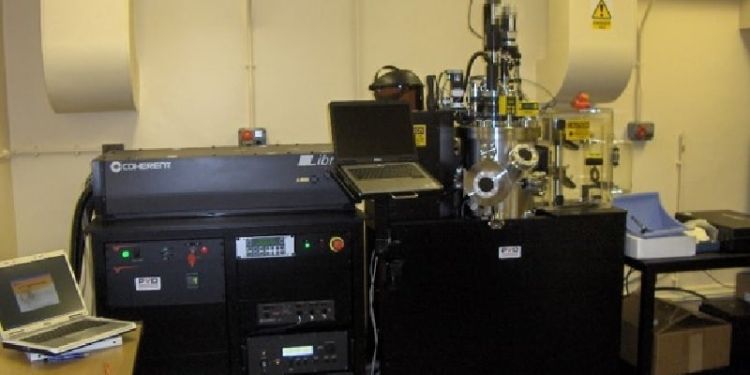
In our experimental approach we investigated deposition of calcium phosphate minerals using ultrafast femto-second pulsed laser deposition of hydroxyapatite on a titanium metal substrate with and without graphene as an electrically conducting medium. The deposited thin film composite materials were analysed for cell proliferation tests to ascertain osteogenic potential for surface modified materials.

Marie Curie Fellowship Training
The project was funded through a Marie-Curie fellowship, which offered opportunity for research training in the areas of advanced bio-materials processing and characterisation, and osteogenic characterisation. The fellowship also provided the opportunity for interaction with Clinician, Prof Giannoudis who is professor and consultant of trauma and orthopaedic surgery. The cell characterisation training was supported by Dr. E M Raif, who trained the MC-fellow in the area of osteoblast cell growth. The materials characterisation support were provided by the Research team in the host institute, and the original research contributions were published in peer-reviewed journal and presented at the conferences.
Research Training Objectives
The main research training objectives for realising the impact of research were:
- To develop an osteoconductive and inductive surface on titanium alloys which is widely used for implant and post-surgery fracture fixation. It is essential that such materials are non-toxic, mechanically robust in the human physiological environment. For this reason, studying the coating of Ti-alloys with osteogenic surface via novel cost-compatible technique will help in developing implant for compromised patients.
- The research training demonstrated that the pulsed laser deposition of HAp films on Ti-alloy and formation of graphene as a compatible surface is possible for surface engineering of implant materials. Such materials were successfully tested for osteo-induction and conduction.
- The research also offered opportunity for bringing together novel tools for materials characterisation and understanding of 2d-materials growth on metal and inorganic glass surface for range of sensing and light-generation device applications, which may be compatible with bio-implants. In this respect, the deposition of 2D-MoS2 materials, as a supplement to graphene and HAp, were studied for light emission properties. The long-term objective is design osteoconductive and inductive bone implants with integrated light-based sensor for monitoring the healing process.

The research training activities which contributed to the overall work programme are briefly described:
- Literature review of the emerging issues on Ti-alloy bone implant integration and current failure rates. It was evident that the patients with 10+-year old implants were more likely to have implant failure due to age, loss of bone stock and implant-bone joint weakening.
- Before the experimental programme could begin the research ethics form was completed as a deliverable and the relevant training for ethics and lab safety were completed. The MC fellow was monitored for progress in meeting his training activities on a fortnightly basis.
- The RTA activities on the following aspects commenced after laser safety course and training were successfully completed.
- Ti-alloy based graphene/calcium phosphate materials processing using fs-PLD: Synthesis of Graphene as strengthening components for bone-template engineering - The first objective was to synthesize graphene using femtosecond pulsed laser deposition (fs-PLD) and grow graphene layers using amorphous carbon films of finite thicknesses and annealing in the presence of metallic nickel (Ni) as a catalyst film. Nickel catalyst was then removed and calcium phosphate layer was grown.
- Characterisation of Graphene and CaP-G on porous Ti-alloys: In the second part of training activities the characterisation of deposited carbon/graphene, Hydroxyapatite materials were carried out using FTIR, X-ray diffraction, and Raman spectroscopy of thin film calcium phosphate minerals and carbon and transformed graphene films. In this training package I also learnt TEM, XPS, and fluorescence spectroscopy of materials.
- Cell cultivation, cytotoxicity, cell attachment and cyto-mineralization: Being a new researcher in biomaterials, I was unfamiliar with cell culture experimental methodologies for which I received training in the Oral Biology lab at the St. James Hospital. The work was supervised by Dr El Mostafa Raif, an academic expert in this field. Once the deposition condition for calcium phosphate and carbon was optimised, the osteoblast related cell culture tests were performed for supporting mineralization. Cell proliferation and viability tests were carried out using laser confocal microscopy and the mineral samples were analysed using the Raman spectroscopy. The effect of nano-scale toxicity was also investigated.
- A new idea was also developed during the MC-project which was whether a 2D-material like MoS2 can also be made compatible on to a glass substrate for measuring bone healing by measuring the characteristics of emitted photons in near-IR. This work led to the growth MoS2 thin films using PLD onto a silica substrate. The photon emission properties were characterised and the results are being further examined for engineering implantable medical device with a light-emitting bone healing sensor.
The research is continuing at the University of Leeds, and is currently supported via a new EU project, led by Prof Peter Giannoudis in the School of Medicine, and three PhD projects at Leeds. The project on bone healing will start in Jan 2020, in which A Jha and Prof Giannoudis are also developing novel sensor methodology for bone healing.The research topic on bone restoration and repair continues to remain quite popular. Currently, via the support of MRC-CiC project, the animal model studies are also being planned. The study will complete in next summer.
Impact
The probability of bone related injury increases with age due to falls, osteoporosis and bone disease. With age, the healing potential of human body also diminishes, which is where the basic research in bone materials is required for promoting bone healing. The data of bone failure from the International Osteoporotic Society is quite compelling in demonstrating the risk of bone failure in 50-60 age group (female and males).
The main cause of bone failure remains due to the loss of bone mineral density for supporting the load-bearing capacity of the body. Combined with the increasing obesity, the bone-failure related morbidity continues increase not only with EU but also in the rest of developed world. The chance of full recovery of a 60+ year old patient suffering from acute osteoporosis [1], after trauma reduces dramatically to less than 25%. Over 300,000 hospital admissions occurred in 2017 [2] due to poor bone stock, as patients were unable to bear fall and injury.
For such bone injury tissue augmentation via increased osteoinduction and conduction is essential for increasing the overall mechanical strength of poor bone stock. It is for this reason, the MC-fellowship research was undertaken, so that a method of enhancing osteoconductivity and inductivity can be increased in vivo, ultimately in bone-compromised patients. A successful project would lead to improved quality of life, low cost of care after surgery, and reduced cost of repeated intervention.
This project has received funding from the European Union’s Horizon 2020 research and innovation programme under grant agreement No 752257
Publications and outputs
Journal Papers
[1] C Maddi, JR Aswin, A Scott, Z Aslam, E Willneff, KNVD Adarsh, A Jha. Structural, Spectroscopic, and Excitonic Dynamic Characterization in Atomically Thin Yb3+‐Doped MoS2, Fabricated by Femtosecond Pulsed Laser Deposition, Advanced Optical Materials, 190075 (2019), online available 5 Sept 2019. https://doi.org/10.1002/adom.201900753.
[2] Yannick Bleu, Florent Bourquard, Teddy Tite, Anne-Sophie Loir, Chirandjeevi Maddi, Christophe Donnet,* and Florence Garrelie. Review of Graphene Growth From a Solid Carbon Source by Pulsed Laser Deposition (PLD); Front Chem. 2018; 6: 572. doi: 10.3389/fchem.2018.00572.
[3] Chiranjeevi Maddi, Florent Bourquard, Vincent Barnier, José Avila, Maria-Carmen Asensio, Teddy Tite, Christophe Donnet & Florence Garrelie; Nano-Architecture of nitrogen-doped graphene films synthesized from a solid CN source; Scientific Reports , (2018) 8:3247, DOI:10.1038/s41598-018-21639-9
Conference Papers / Presentations
[1]. C. Maddi, A. Jha, P. Aparna, KV. Adarsh: Rare-earth Yb-doped MoS2 grown by femtosecond pulsed laser deposition for photonics applications, 8th International Conference on Optical, Optoelectronic, and Photonic Materials and Applications (ICOOPMA-2018), August 28-31st 2018, Maresias-SP, Brazil.
[2]. C. Maddi, P. Aparna, KV. Adarsh, A.J. Scott, A. Jha, Spectroscopic and Structural Properties of Doped and Undoped 2D-MoS2 Thin Films for Optoelectronic and Photonic Device applications, Annual General Meeting of Materials Research Society, Bangalore, India, 12-15 Feb 2019 (Invited Talk).
[3]. C Maddi, JR Aswin, KV Adarsh, AJ Scott, A Jha; Spectroscopic and Structural Properties of Yb3+-Doped and Undoped 2D-MoS2 Thin Films for Optoelectronic and Photonic Device Applications; The European Conference on Lasers and Electro-Optics, cm_p_12 (June 23-27, 2019) Munich CLEO Europe and EQEC).
[4]. M. Chauhan, C. Maddi, A. Jha, V. Subramanian, and P. Valdastri; Characterization of Urease enzyme using Raman and FTIR; Spectroscopy Biophotonics Congress: Optics in the Life Sciences Congress 2019 (OSA), 14-17 April, Tucson, Arizona (Paper: JT4A.46.pdf). Novel Techniques in Microscopy 2019 (ISBN: 978-1-943580-54-5).
[5]. Emaan Alsubhe1, Antonios Anastasiou, Chiranjeevi Maddi, El Mostafa Raif, Peter V. Giannoudis, Animesh Jha, Interaction of Femtosecond Pulsed Lasers with Fe2+ and Fe3+ Doped Calcium Phosphates for Bone Tissue Engineering, Biophotonics Congress: Optics in the Life Sciences Congress 2019 (OSA), 14-17 April, Tucson, Arizona (Paper: JT4A.36.pdf). Published Novel Techniques in Microscopy 2019 (ISBN: 978-1-943580-54-5).