Computer Science in Biology, Medicine and Health
Our research covers a range of applied topics in biology, medicine and health. We construct computational models of biological systems, collaborate with pathologists to make breakthroughs in 3D imaging and digital microscopy, deliver novel designs for bio-inspired autonomous agents, and develop secure systems architectures, data mining and visual analytics methods for Big Data.
We collaborate extensively with the University’s Medicine and Health, Biological Sciences, Chemistry, Mathematics and Engineering departments. We also have substantial collaborations with external organisations such as the Leeds Teaching Hospitals NHS Trust (one of the largest teaching hospitals in Europe) and the Leeds-based NHS Health and Social Care Information Centre. For details, please see our research pages.
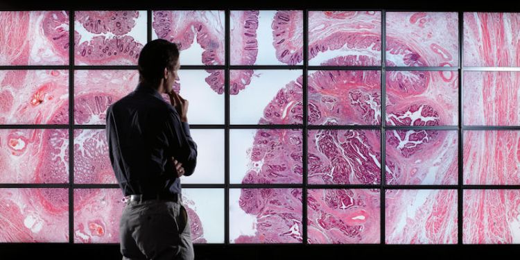
Our facilities include wall-sized visualisation displays and medical-grade high-resolution workstations. We also make substantial use of laboratory facilities in Biological Sciences, and genomics next-generation sequencing and virtual pathology scanning services in Medicine and Health.
We run a regular seminar series every third Thursday at 3:00pm in the School of Computer Science (EC Stoner 6.08).
Importance and impact of our research
Our virtual microscope research aims to develop a visualisation system that out-performs the conventional microscope for cancer diagnosis. A wall-sized version of our virtual microscope is already in use for specialist histopathology training in St James' Hospital. A by-product of our research is that we have developed user interfaces that allow users to efficiently navigate collections of gigantic digital images. The research was one of "100 ground breaking pieces of research" that were featured in Research Council UK's Big Ideas for the Future report.
We have developed software that lets pathologists reconstruct virtual 3D blocks of tissue from hundreds of ultra-thin biopsy slices. After reconstructing the tissue using artificial intelligence techniques, we use 3D computer graphics to let pathologists examining the tissue. Our software has let pathologists understand the structure of cancerous tissue in ways that were impossible with conventional light microscopes.
Further information
View all members of our research group and publications.
PhD projects
We have opportunities for prospective postgraduate researchers. Find out more.
Contact us
If you are interested in collaborating with us or joining our research team, please get in touch with a relevent member of our group.