A sharper focus on protein behaviour
Scientists have developed a new computational technique that allows them to see in finer detail the way protein molecules behave.
Currently, they have relied on laboratory techniques such as X-ray crystallography and cryo-electron microscopy, but those laboratory procedures can disrupt the normal functioning of the molecule.
As an alternative, they could use an atomic force microscope where a sharp probe feels the shape of atoms that are just fractions of a nanometre in size - where a nanometre is one billionth of a metre. But the videos produced by atomic force microscopes have been blurry.
In a scientific paper - Localization atomic force microscopy - published in Nature, researchers have revealed a new way of improving the resolution of the video images.
Understanding proteins key to health research
University of Leeds Alumnus, Dr George Heath, conducted the research in Professor Simon Scheuring's lab at Weill Cornell Medicine in New York before returning to Leeds to build his own research team as a University Academic Fellow.
Dr Heath, based in the School of Physics and Astronomy at Leeds, said: “Atomic force microscopy is a powerful technology that enables scientists to watch proteins in action. But to be able to get the most out of the videos, we have to increase their resolution. That finer focus on the behaviour of proteins is essential if we want to get a better understanding of the way proteins can both prevent and trigger disease.”
Proteins are nature’s workhorses and there are tens of thousands of them in the human body. They come together in fleeting associations to carry out the vital chemical and signalling processes necessary for life.
Super-resolution microscopy
The new technique is based on a concept used in light microscopy called super-resolution microscopy, where light emitted from atoms is used to track their location.
Dr Heath found that by using super-resolution analysis, it was possible to develop a more detailed picture about the structure of proteins.
The new technique can be applied retrospectively to the blurry images captured by an atomic force microscope.
To showcase the power of the method, the researchers generated new high-resolution images of three different proteins including a water channel, a chloride pump and a protein involved in cell membrane repair. Imaging individual molecules as they carry out their functions could open entirely new types of analysis.
Dr Heath will be advancing some of the research findings at the Wolfson Imaging Facility, which is being housed in the new Sir William Henry Bragg building at the University of Leeds.
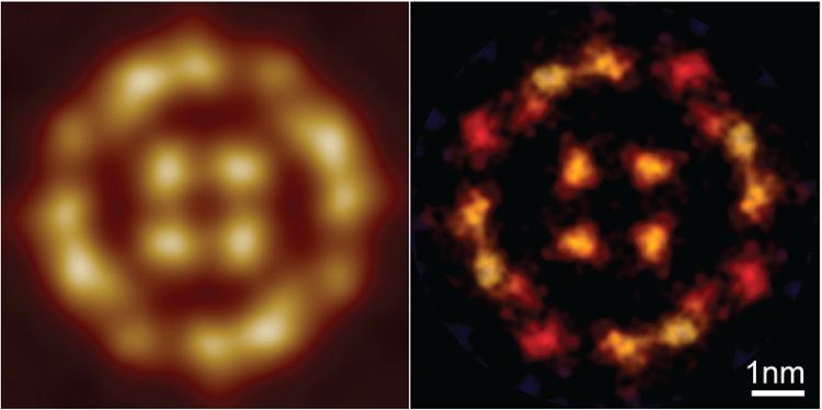
Top image: The picture shows - on the left - an image from a blurry video recorded from an atomic force microscope. The image on the right is from the same set of images which have been processed to build a higher resolution picture.


